X-Ray Absorption Spectroscopy

Products
Fault Isolation
3D X-ray Microscopes
Contact Sales
Products
X-Ray Absorption Spectroscopy
Fault Isolation
3D X-ray Microscopes
Contact Sales
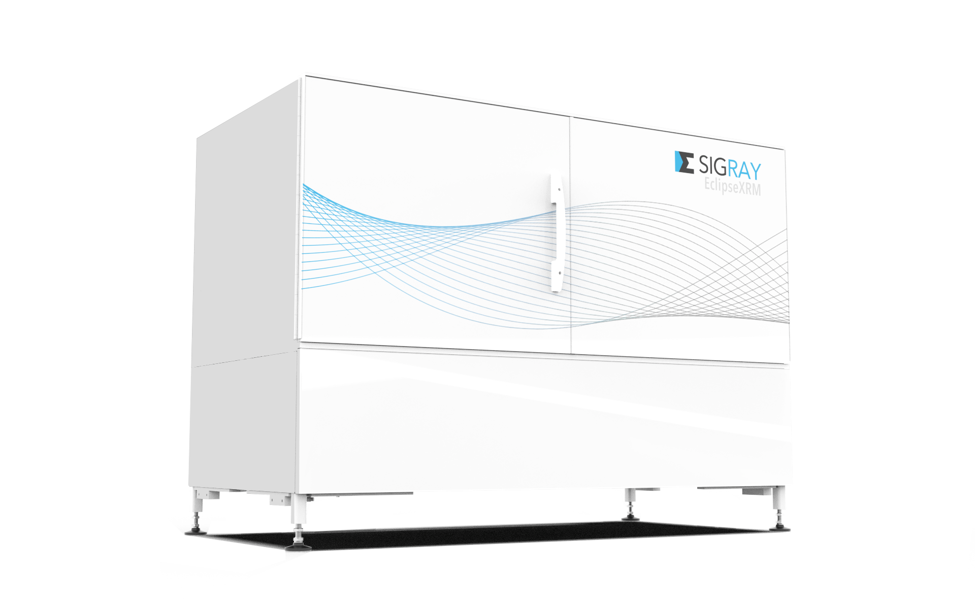
EclipseXRM-910
___________
Highest x-ray resolution performance
Patented technology
Spatial resolution of 0.3 µm
Submicron resolution, even for large working distances
Key Advantages:
- 3D x-ray microscope with breakthrough spatial resolution
Achieve down to 0.3 µm spatial resolution and <30 nm voxels - Maintain high resolution at large working distances
Submicron resolution achieved, even for large source-sample (working) distances. This is important for large or in situ samples - Maximum versatility with novel contrast mechanisms
Patented Multi-Spectral Source (MSS) for quasi-monochromatic illumination to allow up to 10X throughput on the most difficult-to-image, low contrast samples
___________________________________
Leading: 300 nm (0.3 µm) Spatial Resolution
EclipseXRM is a revolutionary system that bridges the x-ray microscope (XRM) gap between micro-XRM and nano-XRM, achieving spatial resolution below 0.3 µm, even for large samples. This far surpasses the leading flexible XRMs on the market, which are limited to ~0.5 µm spatial resolution.
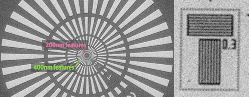
The system achieves the highest resolution in its class using a highly efficient detection system, enabling significantly higher throughput at smaller voxel sizes than other systems on the market.
An example of the EclipseXRM’s resolution advantage over a leading x-ray microscope (XRM) is demonstrated using a shale rock sample (illustrated below). The image on the left shows data from a leading XRM with 0.5 µm spatial resolution using a 20X objective, while the right-hand image showcases the EclipseXRM’s superior high-resolution imaging. Not only is the EclipseXRM data markedly crisper—almost SEM-like in sharpness—but it was also acquired in significantly less time (2.5 hours vs. 4 hours). Furthermore, EclipseXRM’s dataset had sufficient signal-to-noise ratio (SNR) for simple FDK (Feldkamp-Davis-Kress) reconstruction, whereas leading XRM’s 20X data required time-consuming iterative reconstruction due to poor SNR.
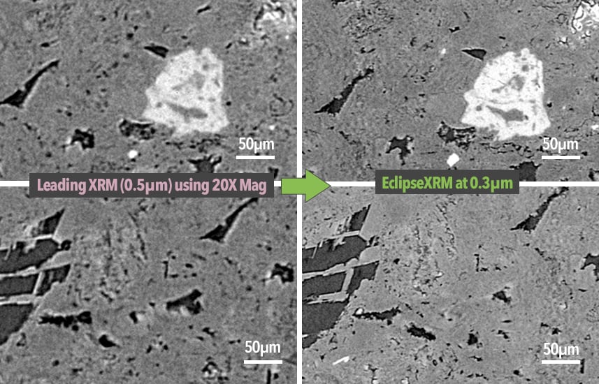
EclipseXRM achieves higher resolution, faster: The same carbonate sample imaged on a leading XRM with two-stage magnification and EclipseXRM. The leading system advertises ~0.5 µm spatial resolution and used a 20X magnification objective to acquire the data in 4 hours, while EclipseXRM acquired 0.3 µm data in <2 hours.
_______________________
Submicron Resolution, even at Large Working Distances
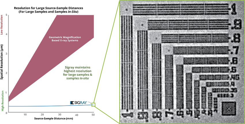
Large samples and in situ samples typically require greater clearance for rotation, which can result in poor resolution on standard x-ray systems. A commonly marketed solution to this issue is a “two-stage magnification” scheme using high-magnification objective lenses. However, this approach compromises detector efficiency.
EclipseXRM’s new, patented architecture provides an alternative, achieving submicron resolution at large working distances without requiring high-magnification objective lenses (see below).
___________________________________
Novel Multi-Spectral Imaging Enables Maximum Versatility
EclipseXRM was engineered to meet the demands of busy central research facilities that handle a wide variety of sample shapes, sizes, and compositions. These facilities are often tasked with achieving the highest image quality in the shortest acquisition times. Since no single set of components can address the full range of needs, EclipseXRM integrates multiple x-ray sources and detectors into a single system. A key option is the Multi-Spectral Source (MSS), which uniquely provides access to multiple quasi-monochromatic x-ray beams—unlike the polychromatic x-rays produced by most system sources. This enables outstanding contrast for extremely challenging samples, such as low-Z structures in novel lithium battery concepts and biological samples.
EclipseXRM’s new, patented architecture provides an alternative, achieving submicron resolution at large working distances without requiring high-magnification objective lenses (see below).
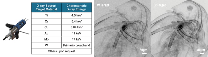
System Features
________________________________
Novel Architecture: Patent-Pending and Patented X-ray Technology
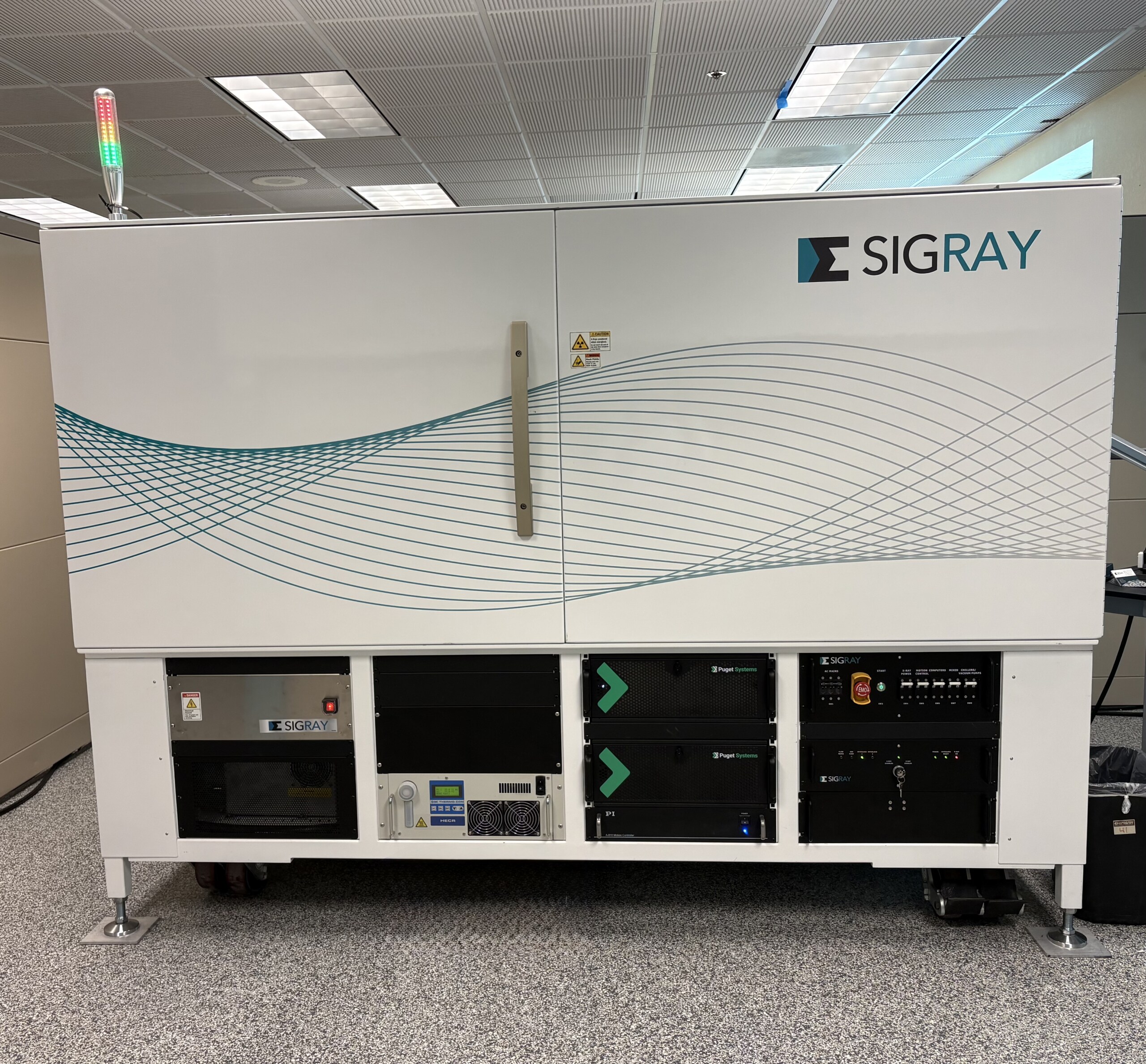
EclipseXRM was designed by Dr. Wenbing Yun, a pioneer in a synchrotron and aboratory X-ray microscopy for over four decades. He has received more than four prestigious R&D 100 awards for his X-ray microscope innovations. The system incorporates several patent-pending and patented technologies developed by Sigray, along with the latest hardware advances, such as maintenance-free air-bearing rotary stages with sub-50nm runout and high-efficiency CMOS sensors.
The system has been adopted by leading semiconductor companies and research institutions worldwide. (To request a complimentary demo on a local system or through Sigray, please fill out the form below).
_____________________________
Progressive Zoom Capabilities
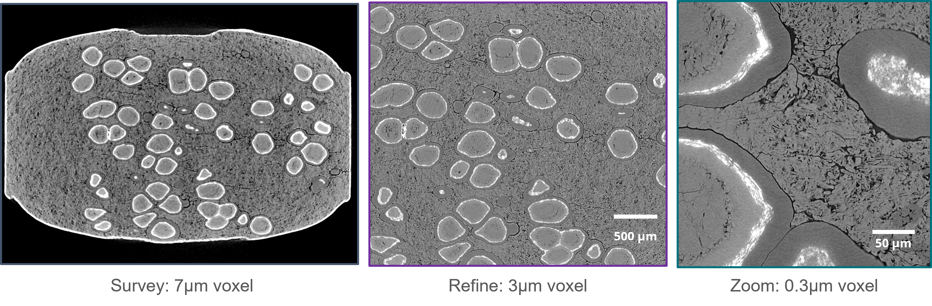
EclipseXRM provides LFOV survey scans (including Ultra-Ultra LFOV scans using a 25MP+ detector) that are used for identifying regions of interest. The regions of interest can be continuously refined without cutting the sample. See below for a commercial anti-acid, progressively zoomed in while keeping the sample intact.
Deep learning based segmentation routine, which improves accuracy and speed of segmentation. Above is a segmentation of different minerals within a kidney stone. Enabled using Dragonfly.
________________________________
Software: GigaRecon Tomography & Sigray3D Acquisition
GigaRecon | A tomographic reconstruction software that delivers the fastest reconstruction times along with an unmatched suite of features for achieving optimal results. Reconstruction speeds of <45 seconds are achieved for 2048 x 2048 x 2200 datasets. GigaRecon also provides the fastest iterative tomography reconstruction on the market, enabling high-quality image reconstruction with five times shorter data collection times, substantially accelerating tomography imaging compared to conventional FDK reconstruction.
Sigray3D | Intuitive Acquisition with XRM Companion
- Easy, intuitive software gets your team up and running in no time
- Click to align the sample and start measuring in seconds
- AI-powered AutoPilot suggests the optimal settings for each sample
- Queue samples with the automated Sample Handling Robot (SHR) for weekend and overnight runs without operator assistance
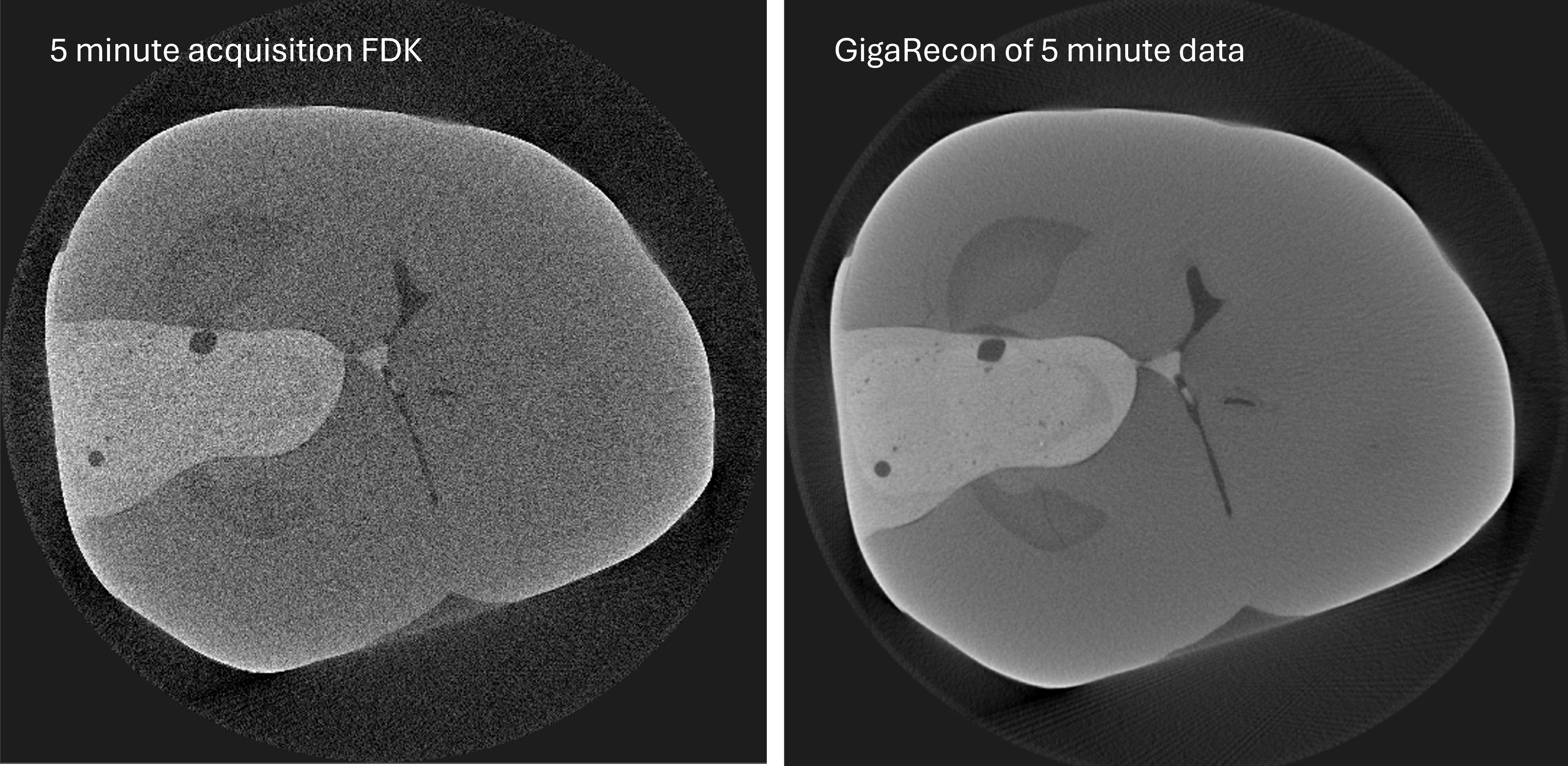
GigaRecon provides powerful reconstruction approaches that can significantly accelerate acquisition time. Left is a human tooth with a metal implant reconstructed through standard FDK (feldkamp), right is Gigarecon reconstruction of the same 5 minute data.
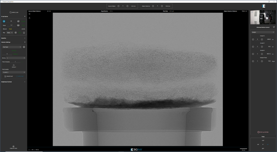
XRM Companion features an intuitive GUI for acquiring and measuring data
_________________________
Phase Retrieval
EclipseXRM provides practical phase contrast for biological, polymeric, and geological samples, accessible on the system without the prolonged acquisition times required by other systems. Additionally, phase contrast information can be easily retrieved using Sigray’s phase retrieval software for quantification and analysis.
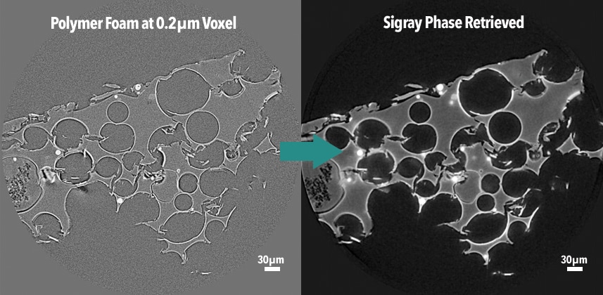
EclipseXRM provides phase contrast imaging capabilities and straightforward phase retrieval to enable enhanced data analysis
_______________________________
Deep Learning / AI Based Routines
Sigray offers a wide variety of AI-enhanced routines, including deep learning to improve segmentation speed (and quality) and to increase acquisition time by 4X or more (see right-hand image).
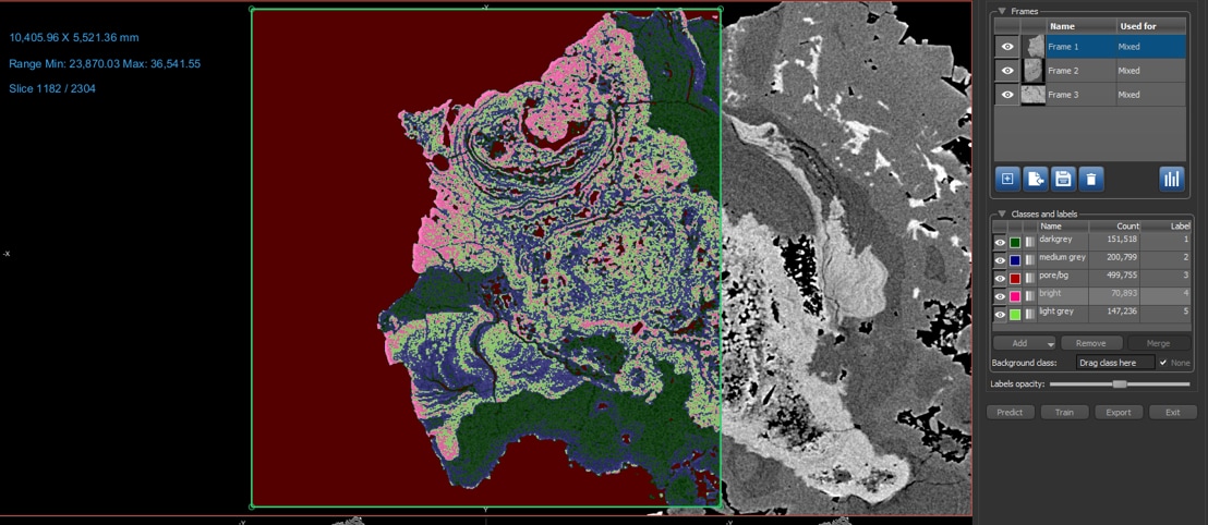
Deep learning based segmentation routine, which improves accuracy and speed of segmentation. Above is a segmentation of different minerals within a kidney stone. Enabled using Dragonfly.
Applications
__________________________
Semiconductor Failure Analysis
3D x-ray microscopy has become the workhorse approach to investigating failures in semiconductor packaging. Due to the ever-decreasing sizes of packaging features, increasing higher resolution is required to detect failures such as cracks, voids, and delamination. The zoomed-in image of a single wire bond shown on the right demonstrates the superior resolution and contrast of EclipseXRM for semiconductor samples.
Note: Most of our results cannot be shown due to NDAs. If you would like a true understanding of our capabilities, please send us a sample so that we can demonstrate them on your sample. The universal response to our semiconductor FA capabilities has been extreme excitement due to the unprecedented level of detail and contrast we achieve.
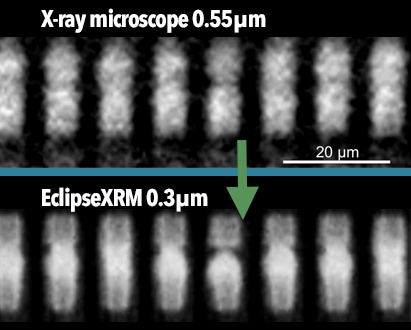
Microbumps imaged: leading XRM vs. EclipseXRM
MicroSD card imaged at 0.15um
______________________________
Intact Batteries and Batteries in-operando
EclipseXRM provides variable resolution within a sample, enabling hierarchical characterization of batteries—from the full field of view (FOV) to detailed region-of-interest imaging—without the need to de-package the battery. This allows for the non-destructive identification of issues such as small defects (e.g., cracks, particles) and shorts. EclipseXRM also delivers high resolution for detailed analysis of electrode particles, capturing their shape, size, and any defects (e.g., voids, cracks).
Sigray’s powerful capabilities for batteries and the associated papers can be found on its Batteries applications page.
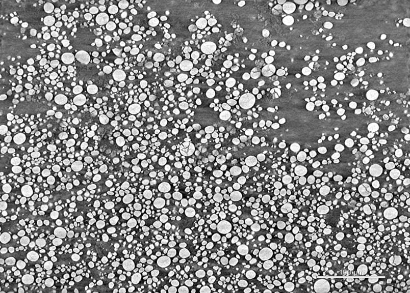
Ultrahigh resolution (0.21 um voxel) imaging of battery cathode particles
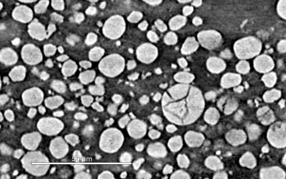
Region of interest of battery cathode particles (0.21 um)
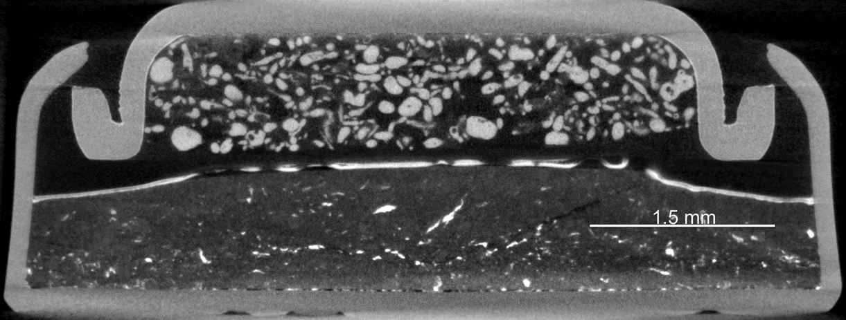
Cross-sectional view of an intact coin cell, showing flexibility of EclipseXRM
____________________________
Biological and Polymer Samples
Biological and other low-density samples, such as polymers, can be extremely challenging to image using conventional x-ray techniques like microCT due to their low absorption and ultrahigh resolution requirements. EclipseXRM provides both superior contrast and resolution, enabling the visualization of sub-cellular details.
Shown in the example to the right is a mouse sciatic nerve sample, demonstrating how the system’s ultrahigh resolution clearly reveals the axons and myelin sheaths.
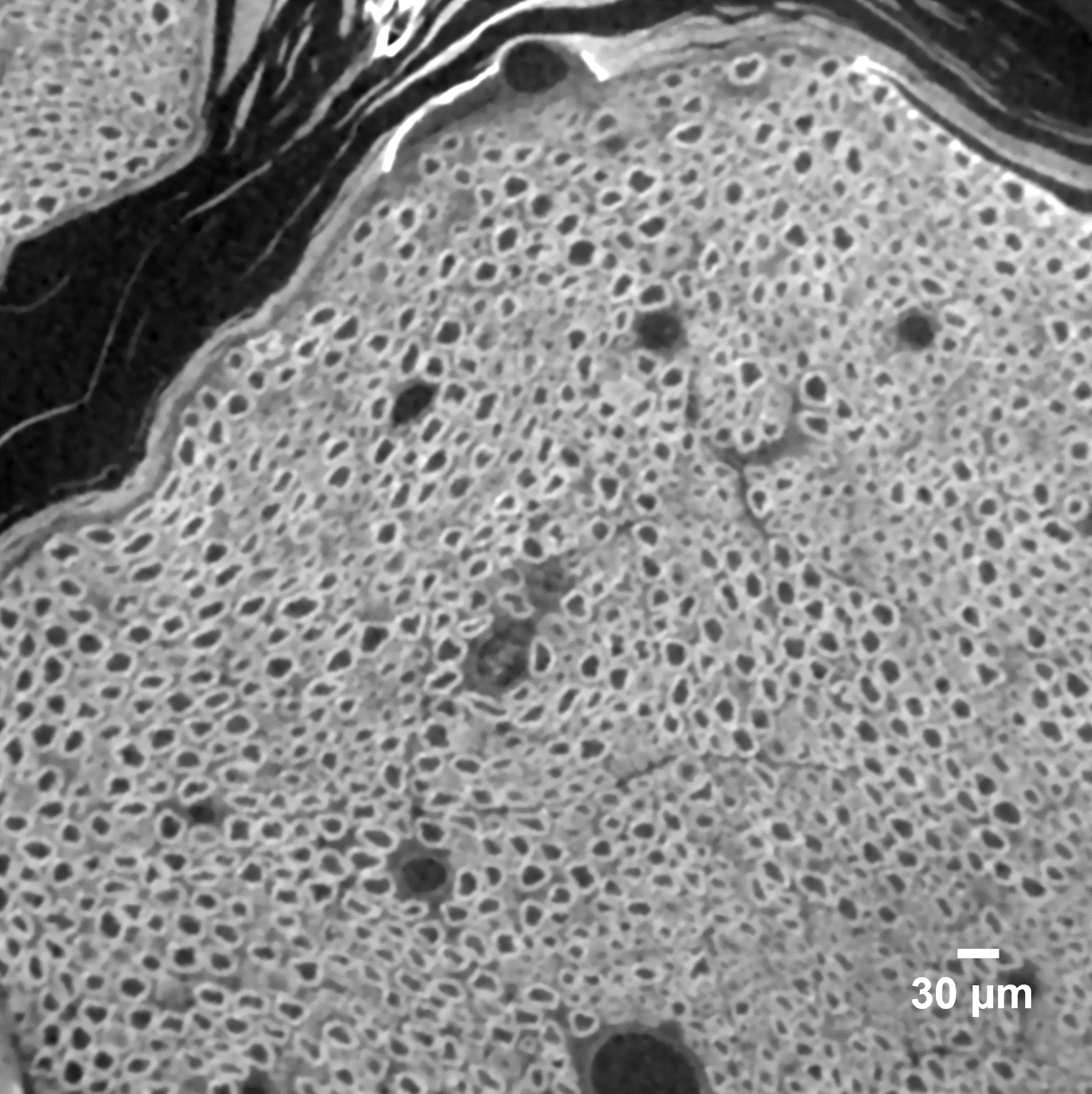
Mouse sciatic nerve samples at 0.25 µm
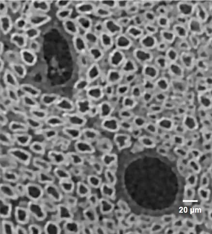
Cropped region of interest of mouse sciatic nerve cells (0.25 µm)
___________________________
In-situ Microstructural Evolution
A significant advantage of EclipseXRM’s design is its ability to maintain high submicron resolution for large samples, enabling high fidelity imaging of specimens placed in larger in-situ cells. The system can capture 3D micro-structural evolution in samples under various conditions, including heating, cooling, tension and compression (T&C), fluid/gas flow, and more. We provide robust in-situ solutions tailored to your needs.
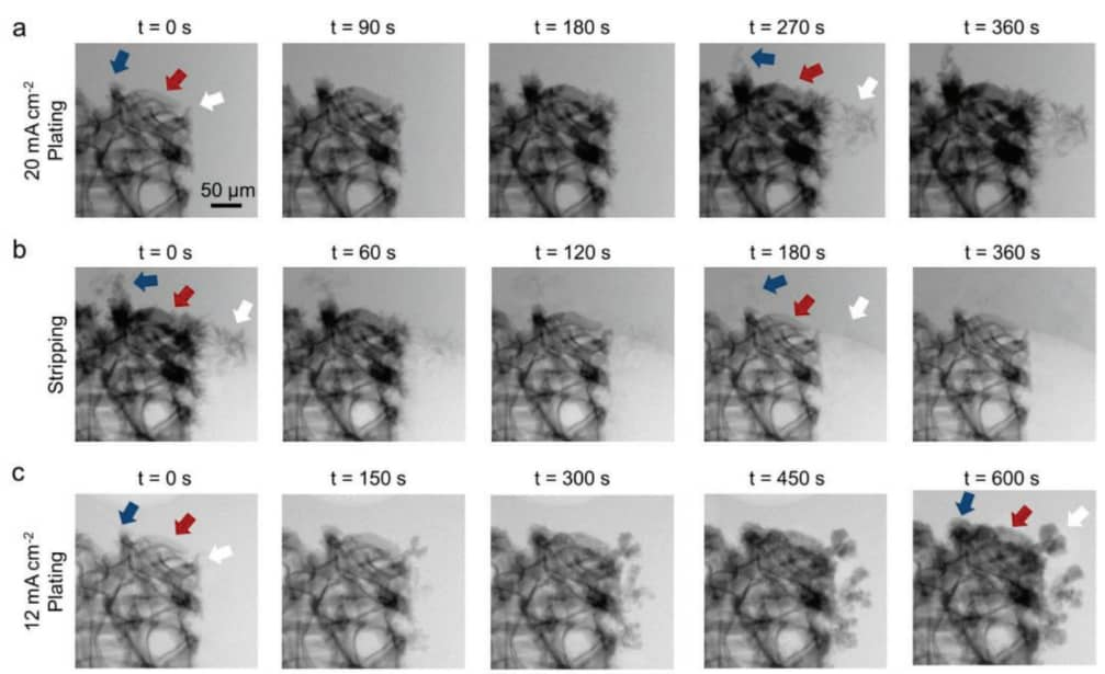
Dendritic growth imaged on PrismaXRM (Sigray’s leading XRM prior to EclipseXRM’s release) on a Zn-ion battery.
Ref: Qian, G., Zan, G., Li, J., Lee, S.-J., Wang, Y., Zhu, Y., Gul, S., Vine, D. J., Lewis, S., Yun, W., Ma, Z.-F., Pianetta, P., Lee, J.-S., Li, L., Liu, Y., Structural, Dynamic, and Chemical Complexities in Zinc Anode of an Operating Aqueous Zn-Ion Battery. Adv. Energy Mater. 2022, 12, 2200255.
https://doi.org/10.1002/aenm.202200255
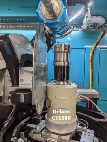
In-situ cells are manufactured by a variety of manufacturers and can provide heating, T&C, fluid flow, and electrical power
Technical Specifications of the EclipseXRM-910
|
|
Parameter |
Specification |
||
|---|---|---|---|---|
|
Overall |
Spatial Resolution |
0.3 µm |
||
|
Resolution at Large Working Distances |
Submicron |
|||
|
Contrast |
Nanofocus X-ray source |
|||
|
Source |
|
|
||
|
Source 2 (Optional) |
160 kV |
|||
|
Detector(s) |
Types |
Two detectors provided standard |
||
|
Detector 1 |
HyperCapture - Standard Flat Panel detector: 7MP with 50 µm pixels |
|||
|
Detector 2 |
|
|||
|
Software |
Command and Control |
Sigray 3D |
||
|
Reconstruction |
GigaRecon - fastest commercial CBCT reconstruction software (ask for our white paper) |
|||
|
Offset Scans |
Expands the horizontal FOV for ultrawide samples |
|||
|
Helical Scan |
Option enabled for tall, cylindrical samples |
|||
|
AutoPilot |
AI-assisted microscope operation for unsupervised acquisition |
|||
|
Linux Workstation |
Interface is on a Windows workstation, while a separate robust Linux workstation controls the system. Advantageous for reliable 24-7 operation. |
|||
|
EPICS |
Open-source software controls for maximum flexibility and software extendibility |
|||
|
Dimensions and System |
Footprint |
|
||
|
Stages |
|